Lower Leg Bone Diagram : Bones of the lower leg : Click now to learn more about the bones, muscles, and soft tissues of these regions at kenhub!
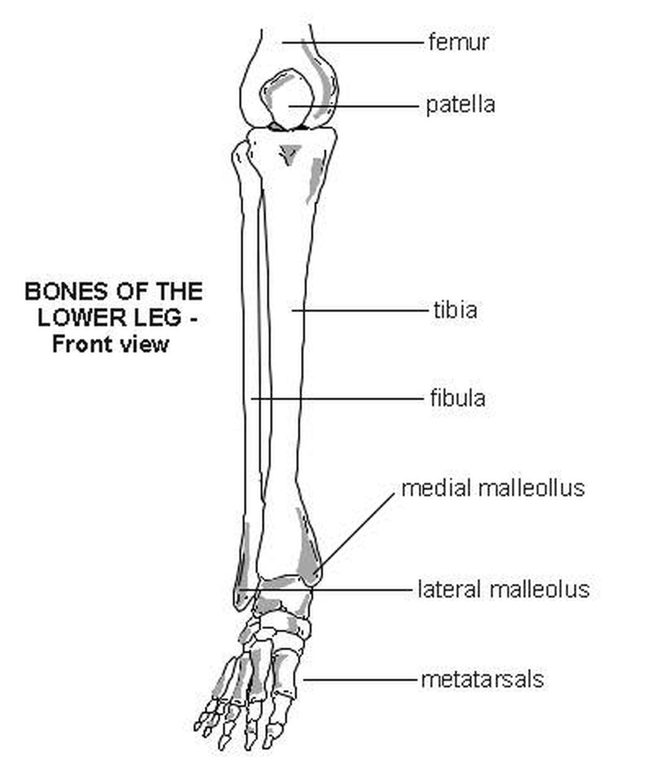
Lower Leg Bone Diagram : Bones of the lower leg : Click now to learn more about the bones, muscles, and soft tissues of these regions at kenhub!. The thigh bone, or femur, is the large upper leg bone that connects the lower leg bones (knee joint) to the pelvic bone (hip joint). Ankle and foot bones and joints unit 4/12/18 lower leg: Related posts of bone anatomy lower leg. The larger bone we refer to as the tibia and is present in front of the lower leg. While their parts are similar in general, their structure has been adapted to differing functions.
Lower jaw (mandible) collar bone. Cheek bone (zygoma) upper jaw (maxilla). Leg picture image on medicinenet com. Frontal, medial/lateral, dorsal, cruciate bursae. 2006 kia optima belt diagram.
Pictures Of Bones Of The Lower Extremities from healthiack.com Common fibula fracture sites are: The knee joint is the largest joint in the body and is primarily a hinge joint, although some sliding and rotation occur. Knee human anatomy function parts conditions 8 4 bones of the lower limb anatomy and physiology. The two bones beneath your knee that make up your shin are your tibia and fibula. The lower leg contains two major long bones, the tibia and the fibula, which are both very strong skeletal structures. The lower leg has a structure by two bones. Vtt 150 horse leg anatomy diagram quizlet. The primary cells in this area are termed as the calf.
When you stand or walk, all the weight of your upper body rests on them.
5 powerful lower body strength routines. 2006 kia optima belt diagram. The thigh bone, or femur, is the large upper leg bone that connects the lower leg bones (knee joint) to the pelvic bone (hip joint). Moreover, the fibula is the smaller bone that goes towards the back part of the leg. Posted on april 18, 2019april 18, 2019. Download a free preview or high quality adobe illustrator ai, eps, pdf and high resolution jpeg versions. Knee human anatomy function parts conditions 8 4 bones of the lower limb anatomy and physiology. The foot bones shown in this diagram are the talus, navicular, cuneiform, cuboid, metatarsals and calcaneus. The primary cells in this area are termed as the calf. Ankle and foot bones and joints unit 4/12/18 lower leg: We think this is the most useful anatomy picture that you need. Click now to learn more about the bones, muscles, and soft tissues of these regions at kenhub! The human body is the structure of a human being.
Download a free preview or high quality adobe illustrator ai, eps, pdf and high resolution jpeg versions. Bones of the human body diagram. Diagram femur bone diagram data pre. Bones lower anatomy limb skeletal human system diagrams leg bone labeled diagram skeleton physiology extremities hand upper jb004 k12 sd template. Leg picture image on medicinenet com.
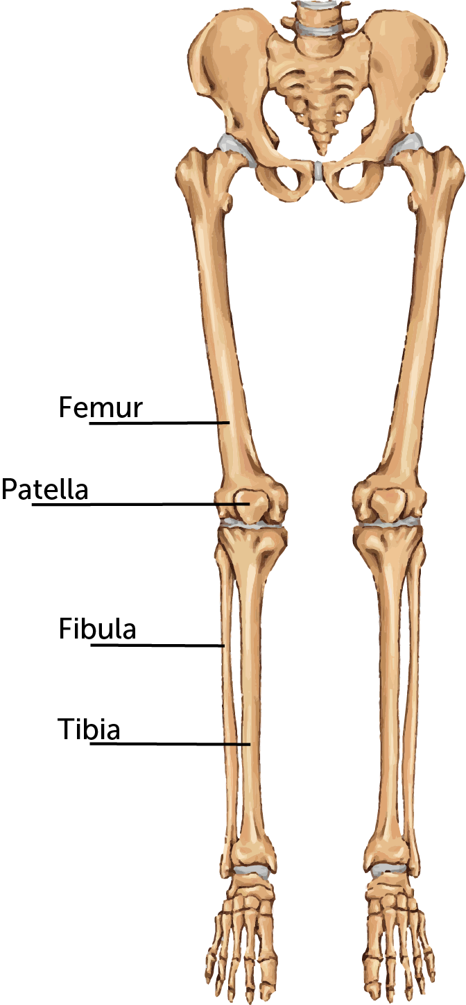
Clipart skeleton broken, Clipart skeleton broken ... from webstockreview.net Knee human anatomy function parts conditions treatments. The bones of the leg are the femur, tibia, fibula and patella. Like the upper limb, the lower limb is divided into three regions. The human leg, in the general word sense, is the entire lower limb of the human body, including the foot, thigh and even the hip or gluteal region. The human body is the structure of a human being. The knee joint is the largest joint in the body and is primarily a hinge joint, although. And the calf is actually a group of various. Bones of the human body diagram.
Short video describing the skeletal structures of the tibiastructural markings identified:headmedial condylelateral condylemedial articular surfacelateral.
The two bones beneath your knee that make up your shin are your tibia and fibula. Your leg bones are the longest and strongest bones in your body. Horse leg bones diagram quizlet. The lower leg is comprised of two bones, the tibia and the smaller fibula. Anatomy leg bone lower foot calf diagram human bones ankle joint system poster muscles fpnotebook parts ortho muscle restrictions printing. Learn vocabulary, terms and more with flashcards, games and other study tools. Diagram femur bone diagram data pre. The lower leg is a major anatomical part of the skeletal system. Like the upper limb, the lower limb is divided into three regions. This lesson gives detailed information of the location diagram of skeletal muscle tissue. Vector illustration with human skeleton scheme isolated on a white background. While their parts are similar in general, their structure has been adapted to differing functions. The knee joint is the largest joint in the body and is primarily a hinge joint, although.
Vtt 150 horse leg anatomy diagram quizlet. The tibia (shin bone) is the medial bone of the leg and is larger than the fibula, with which it is paired (figure 3). Bones lower anatomy limb skeletal human system diagrams leg bone labeled diagram skeleton physiology extremities hand upper jb004 k12 sd template. Learn vocabulary, terms and more with flashcards, games and other study tools. The foot bones shown in this diagram are the talus, navicular, cuneiform, cuboid, metatarsals and calcaneus.
labeled muscles of lower leg - Yahoo Search Results ... from i.pinimg.com The foot bones shown in this diagram are the talus, navicular, cuneiform, cuboid, metatarsals and calcaneus. And the calf is actually a group of various. The human leg, in the general word sense, is the entire lower limb of the human body, including the foot, thigh and even the hip or gluteal region. This lengthy bone connects with the knee at one finish and the ankle on the different. The human body is the structure of a human being. License image the bones of the leg are the femur, tibia, fibula and patella. Learn vocabulary, terms and more with flashcards, games and other study tools. This diagram depicts lower leg bones 1024×1350.
The foot bones shown in this diagram are the talus, navicular, cuneiform, cuboid, metatarsals and calcaneus.
Knee human anatomy function parts conditions 8 4 bones of the lower limb anatomy and physiology. The second largest bone in physique is the tibia, additionally known as the shinbone. The larger bone we refer to as the tibia and is present in front of the lower leg. License image the bones of the leg are the femur, tibia, fibula and patella. Start studying lower leg bone structure. Vtt 150 horse leg anatomy diagram quizlet. 2006 kia optima belt diagram. Frontal, medial/lateral, dorsal, cruciate bursae. Your upper and lower leg are connected by a hinge joint. The two bones beneath your knee that make up your shin are your tibia and fibula. The foot bones shown in this diagram are the talus, navicular, cuneiform, cuboid, metatarsals and calcaneus. The tibia (also called the shinbone) is located near the midline of. Related posts of bone anatomy lower leg.
2006 kia optima belt diagram leg bone diagram. Horse leg bones diagram quizlet.